Dr. Mary E. Morrison
Assistant Professor, Biology Department, Lycoming College
700 College Place, Williamsport, PA 17701
office phone (570) 321-4184
email
Morrison@lycoming.edu
|
Course Syllabi: |
|
|
Bio 447 Cell and Molecular Biology
Research Methods Fall 2009 |
|
Useful links: |
|
Research Summary:
Dr. Morrison was the recipient of a National Science Foundation Support of Mentors and Students (SOMAS) award for summer 2006 research with undergraduates.
Areas of interest include cell biology, neuroscience, developmental biology, virology, and biology education for nonmajors. Possible research projects for undergraduates include:
1) Cell biology of the brain: Granule neuron regulation of Purkinje cell development
2) Molecular biology of the brain: Gene expression changes and signaling events during development of the cerebellum
3) Mammalian Genetics: effects of mutations on mouse brain development
4) Biotechnology: developing methods for gene transfer into neurons
|
Images from research projects: |
|
|
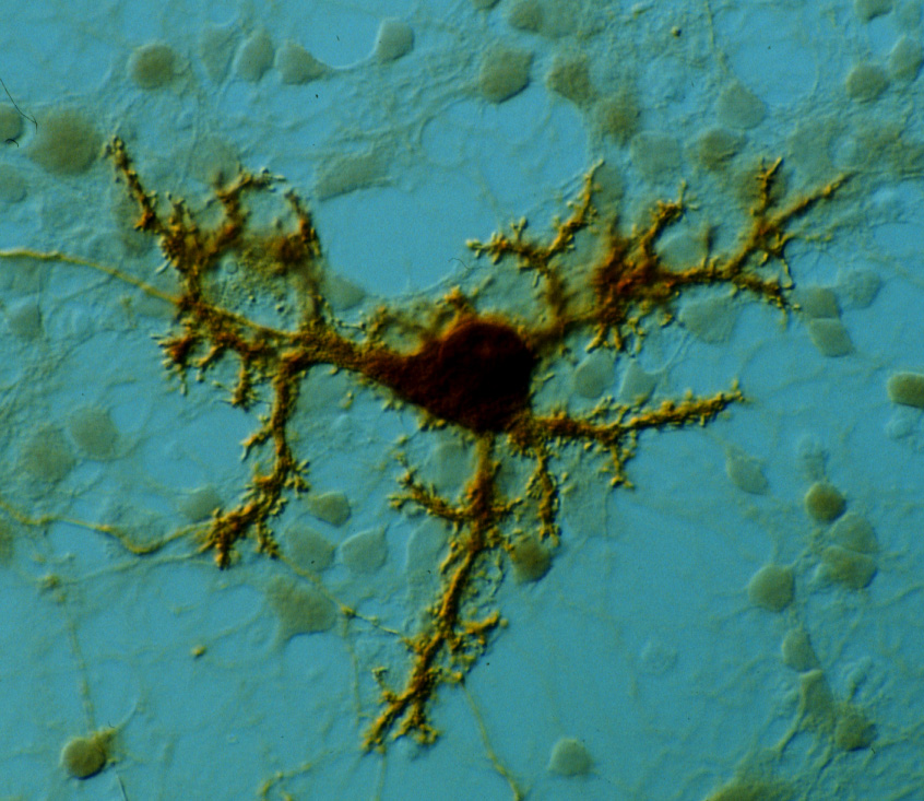
Purkinje cell (brown) cultured with granule neurons for 14 days; stained with anti-Calbindin-D28k/peroxidase. Note the branching dendrites and numerous small, thornlike spines. |
Purkinje and granule cells cocultured for 4 days; stained with: anti-Calbindin-D28k/Cy3 (red), anti-BrdU/FITC (green), and Hoechst 33258 (blue). |
|
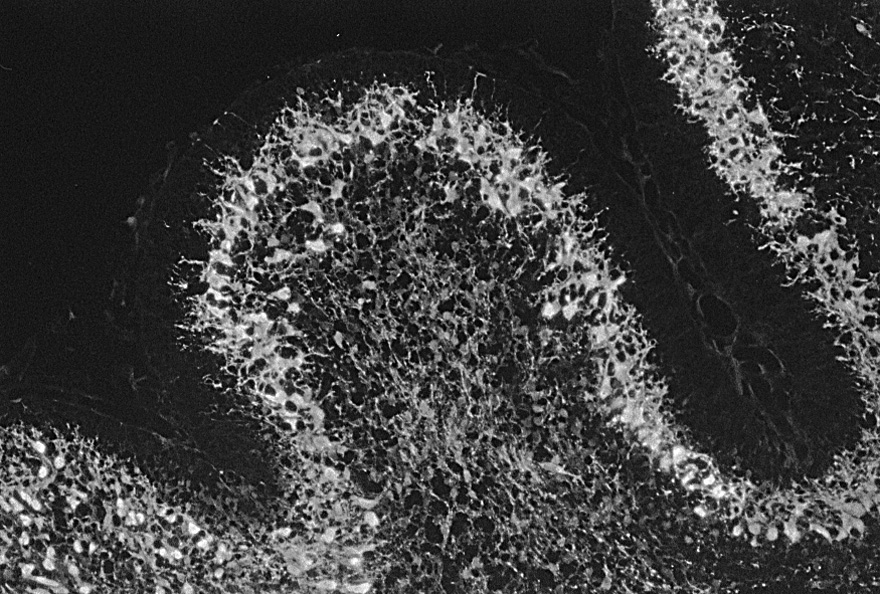
Rat cerebellum, postnatal day 1, Purkinje cells stained with anti-Calbindin-D28k. Note small cell bodies and generally disorganized Purkinje cell layer. |
Rat cerebellum, postnatal day 5, Purkinje cells stained with anti-Calbindin-D28k. Note enlarging cell bodies and Purkinje cell apical dendrites pointing toward the surface of the brain. |
|
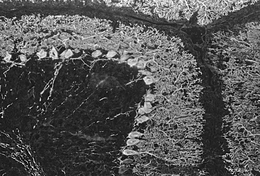
Rat cerebellum, postnatal day 10, Purkinje cells stained with anti-Calbindin-D28k. Note elaborate dendritic trees and Purkinje cells lined up in a monolayer. |
Rat cerebellum, postnatal day 15, Purkinje cells stained with anti-Calbindin-D28k. Note mature configuration of Purkinje cells in a monolayer, with highly branched dendrites. |
|
MRC-5 lung fibroblast cells stained with anti-vimentin antibody/Alexa 488, by AJ Willouer for a Cell Biology class research project |
HeLa human cervical carcinoma cells stained with anti-vimentin antibody/Alexa 488, by AJ Willouer for a Cell Biology class research project |


|
Images outside the classroom: |
|
|
|
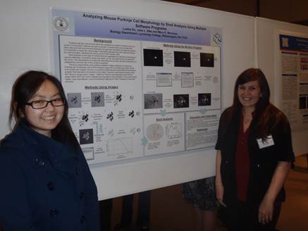
Lusha Xu ’11 and Julia Was ’10 presenting their research poster on
dendrite morphology analysis at the 2011 Lehigh Valley Society for
Neuroscience meeting at Lafayette College. |
John Mastrobuono ‘11 presenting his research poster on
early-stage Purkinje cell markers at the 2011 Lehigh Valley Society for
Neuroscience meeting at Lafayette College. |
Ethan
Sellers ’12 presenting his research poster on early-stage Purkinje cell
markers at the 2011 Lehigh Valley Society for Neuroscience Meeting at
Lafayette College. |
|
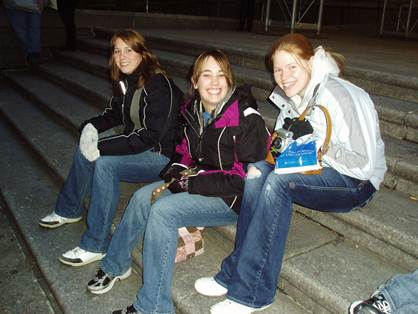
Intro Bio students resting up during the annual trip to the American
Museum of Natural History in New York |
|
|
|
|
|
|
|
|
|
|


|
Images from the Lycoming College Biology
Department |
|
|
|
The spring 2011 Bio 337 Neurobiology class using their
brains! |
KC Failor ’11 and Julia Was ’12 giving their final project
presentation in Bio 435 Cell Biology class |
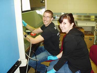
KC Failor ’11 and Julia Was ’12 culturing HeLa cells for
Cell Biology class in our BSL2 tissue culture lab |
|
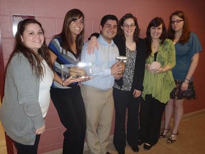
The fall 2010 Cell Biology class all dressed up |
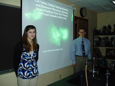
Lauren Bottorf ’12 and Jordan Krebs ’13 giving their final
project presentation in Bio 435 Cell Biology class |
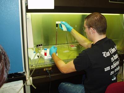
KC Failor ’11 culturing HeLa cells in the BSL2 hood for
Cell Biology class |
|
|
|
|
|
Stamatis Zeris '06, independent study student, using the Nikon microscope |
Mary Gantz '06 and Ashley Campbell '08 working in Dr. Morrison's prep room |
Betsy Reese '07 and Cynthia Smith '07 purifying white blood cells with Ficoll gradients in Bio 347 Immunology lab |
|
Mary Gantz '06 on the Nikon microscope |
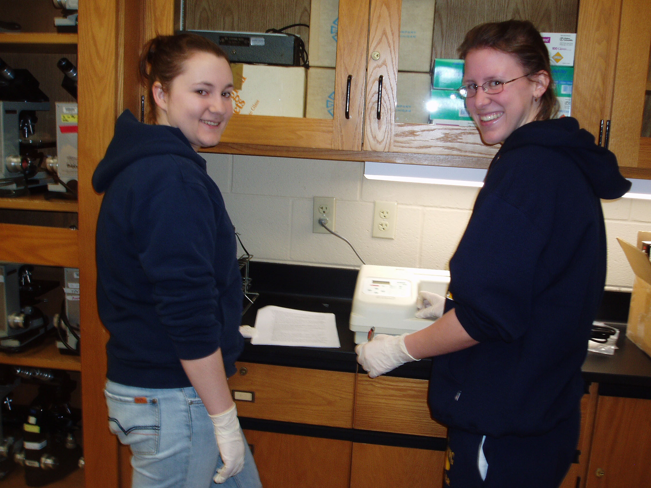
Sara Gavlock '07 and Jessica Bennett '08 programming the microplate reader in Bio 347 Immunology lab |
Bio 106 students celebrating the end of their fermentation lab experiment |